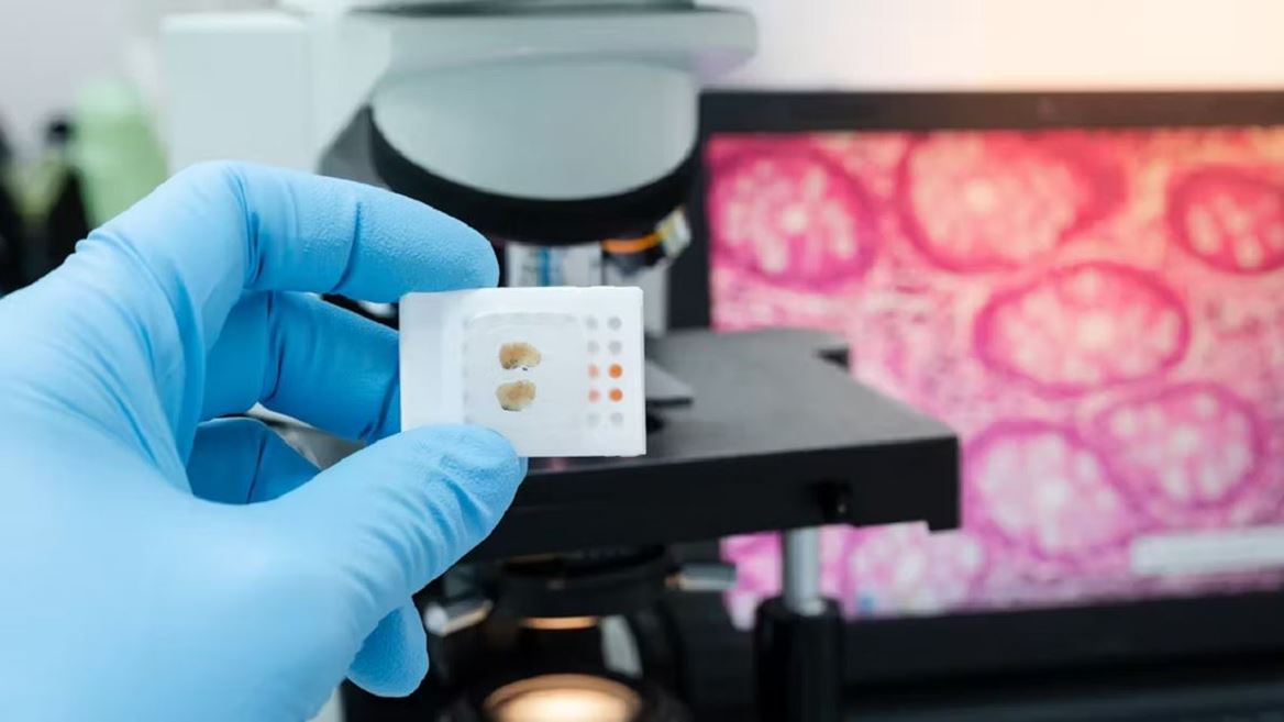
Precision Tissue & Cellular Diagnostics – Histopathology & Cytology
Blufrog Path Lab Solutions, LLC provides comprehensive histopathology and cytology services, delivering accurate diagnoses from tissue biopsies, surgical specimens, and cellular samples. Our CAP-accredited laboratory uses advanced tissue processing, microtomy, routine and special stains, immunohistochemistry (IHC), and in situ hybridization (ISH) to characterize neoplastic and non-neoplastic diseases.
Our board-certified pathologists provide detailed, clinically actionable reports for surgical resections, core needle biopsies, endoscopic biopsies, and frozen sections. In cytology, we offer expertise in gynecological cytology (Pap smears), non‑gynecological exfoliative cytology (urine, effusions, respiratory), and fine‑needle aspiration (FNA) with rapid on‑site evaluation. Digital pathology integration allows remote consultations and second opinions from sub‑specialists.
Gold-Standard Histology
Fully automated tissue processors, high‑quality embedding, and microtomy ensure optimal sectioning for accurate morphological assessment.
Rapid Turnaround
Routine biopsies reported within 48–72 hours; frozen sections within 20 minutes. STAT requests prioritized for critical cases.
Expert Pathologists
Sub‑specialized in breast, GI, GU, gynecologic, pulmonary, and head/neck pathology. Dedicated cytopathologists for FNA and fluid specimens.
Integrated Diagnostics
Seamless correlation with molecular, immunohistochemical, and cytogenetic results for comprehensive patient management.
Need help with a pathology case?
Our expert pathologists are available for consultations and second opinions.
Contact Us Now